South Australian Medical Heritage Society Inc
Website for the Virtual Museum
Home
Coming meetings
Past meetings
About the Society
Main Galleries
Medicine
Surgery
Anaesthesia
X-rays
Hospitals,other organisations
Individuals of note
Small Galleries
Ethnic medicine
- Aboriginal
- Chinese
- Mediterran
Coulter Counter & Coulter Principle
Acknowledgments: We are grateful to Drs. Anne Hamilton-Bruce and Austin Milton who allowed us to photograph some of the old machines in the Basil Hetzel Research Institute and to Dr. John Norman and George Paczek, the senior scientist in the QEH Department of Haematology, who showed us the current model of a cell counter based on the Coulter Principal.
Fifty-six years ago Wallace and Joseph Coulter, working in a Chicago basement. developed a novel way of counting microscopic particles subsequently recognised as the “Coulter Principle”. Their work was patented in 1953 and brought automation to Haematological laboratories. The counting chambers and manual counting became obsolete. (See appendix).
Numerous modifications and improvements produced the current models which produce a record of haemoglobin, haematocrit, red and white cell numbers as well as platelets from a 100uL sample of blood within minutes (commonly known as a “FBC” full blood count). The principle involves a small pore, ie. a microscopic opening separating two volumes of an electrolyte solution. As particles pass across the pore their surface charge alters the resistance across the aperture and this can be recorded. The change in resistance is proportional to the particle size and surface charge and as various blood cells differ in size and surface charge, their numbers and types can be recorded.
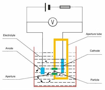
Sketch of the Coulter principle from the home page of Pulver Technology
http.//folk.uio.no/anderne/research html
In subsequent years the Coulter principle was adopted by other companies such as Beckman, Synmex and others and it is now possible to record up to 36 parameters relating to various types of blood cells.
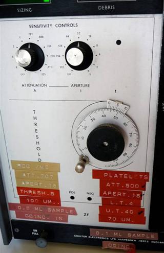
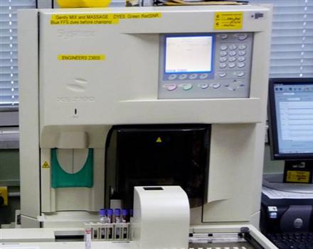
The control panel of an early Coulter counter used in the Department of Medicine of the QEH in the 1970s. Unlike the current automated models the threshold, aperture, and attenuation had to be set manually. The stickers indicate the settings for counting of platelets.
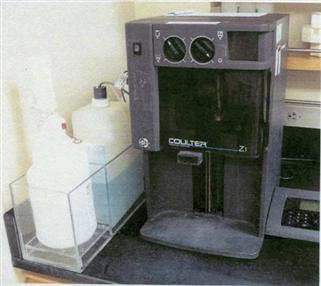
Later model of a Coulter counter showing the apparatus (centre), digital controls (right), and rinsing and diluting solutions (left).
Fully automated counter produced by Synmex Medical Instrument Co.
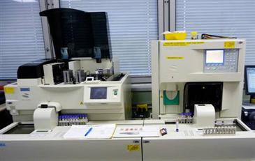
The cell counter is on the right. The machine, which produces and stains blood films, is on the left.
Close up of the cell counter. The blue topped vials containing the blood samples are on the left. The machine pierces the tops, aspirates and dilutes the samples, and produces a print-out record.
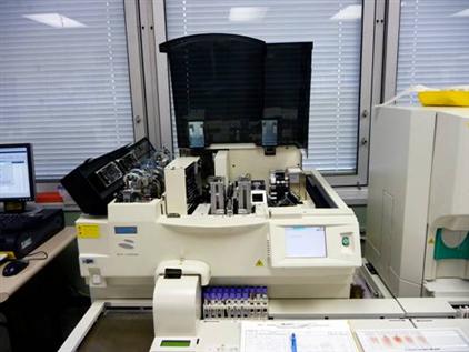
Close up view of the slide machine. The blue-top samples are in the centre. The machine prepares the slides (right) and stains them (left).
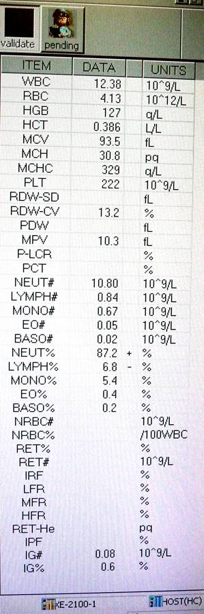
Printout of 36 parameters related to sizes, types & contents of blood cells
.....
- HGB 127 q/L
- HCT 0.386 L/L
- MCV 93 5 fL
- MCH 30.8 pq
- MCHC 329 q/L
- F'LT 222 10^9/L
- POW-SD fL
- RDW-CV 13.2 %
- PDW fL
- MPV 10 3 fL
P-LCR - POT
- NELIT# 10.80 10 ^ 9/1_
- LYMPH# 0.84 10^9/L
- MONO# 0.67 10^9/L
- E0# 0 05 10^9/L
- BASO# 13 02 10^9/L
- NEUT% 87.2 +
- LY1. APH % 6.8 - %
- f.A0NO% 5.4 %
- EO% 0.4
- BASO% 0 2
- NRBO# 10"9/L
- NRBC% /100WBC
- PET%
- RET# 10^9/L
- IRF
....
APPENDIX: Manual counting before Coulter automation
Haemocytometer
OWNER: Dr. Richard Bowering PURPOSE: Counting of red and white blood cells. DESCRIPTION: Red box 20x 8 cm. labelled “Haemocytometer”. CONTENTS: Counting chamber, cover slide, red and white mixing pipettes, & fluids.
Before the advent of the Coulter counter the estimation of the red and white blood cells was done manually with the aid of a microscope.
The counting chamber had engraved counting nets and the number of blood cells per square was related to the total blood count. Six or more models of counting chambers were produced.
A typical chamber had a depth of 1/10 mm and 400 squares with 1/10 mm sides giving a volume of 1/4000 th cubic mm. A red mixing pipette was used for the red cell count, and the white pipette for white cells. A different dilution fluid was used for red or white cells.
Blood was sucked up to the 0.5 or 1.0 mark on the pipette giving a dilution of 1/200 or 1/100 when the appropriate diluting fluid is aspirated to the 101 mark and thoroughly mixed. The counting chamber is then filled.
Usually one counts the number of cells over 5 large squares (5 x 16 ie 80 small ones). The number of small squares counted is then multiplied by the volume (4,000) and the dilution (200) and divided by the number of small squares counted (80). If the total number of red cells counted is 500 then the red blood cell count will be 500 x 200 x 4,000 divided by 80 = 5,000,000 red cells per cubic mm. A similar formula was used for determining the white cell count.
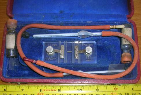
A Buerker counting chamber, red and white pipettes and diluting fluid
For other haemocytometers, click here
-o0o-